
 |
Clinical Image
Central nervous system infection with pyogenic ventriculitis
1 Internal Medicine Resident, Department of Internal Medicine, Unidade Local de Saúde de Braga, Braga, Portugal
2 Internal Medicine Physician, Department of Internal Medicine, Unidade Local de Saúde de Braga, Braga, Portugal
Address correspondence to:
Rui Jorge Silva
Sete Fontes – São Victor, 4710-243 Braga,
Portugal
Message to Corresponding Author
Article ID: 100086Z09RS2026
Access full text article on other devices

Access PDF of article on other devices


How to cite this article
Silva RJ, Antunes E, Silva I, Caridade S. Central nervous system infection with pyogenic ventriculitis. J Case Rep Images Med 2026;12(2):1–3.ABSTRACT
No Abstract
Keywords: Central nervous system infection, Magnetic resonance imaging, Pyogenic ventriculitis
Case Report
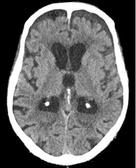
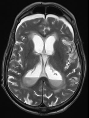
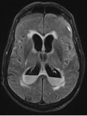
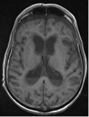
An 89-year-old man, with a history of chronic kidney disease (enrolled in a regular hemodialysis program) and early-stage dementia, presented to the emergency department with symptoms that began one day before admission, including fever, sweating, shivering, dyspnea, productive cough, and disorientation. Approximately 2 weeks earlier, the patient sustained a fall resulting in a frontal traumatic brain injury, with a forehead laceration requiring 8 sutures. At that time, a cranial computed tomography (CT) scan revealed chronic subdural hematomas and a small left frontal subarachnoid hemorrhage, and the patient was subsequently discharged. During the current emergency department visit, he was diagnosed with bilateral community-acquired pneumonia complicated by type 1 respiratory failure. He was hospitalized and was started on empirical antibiotic therapy (ceftriaxone and clarithromycin). On the second day of hospitalization, the patient’s altered mental status worsened; he became nonverbal and localized to pain with the left upper limb. Over the course of a few hours, he rapidly deteriorated to deep coma with absent response to painful stimuli. A CT scan of the brain was performed, revealing cerebral atrophy, bilateral frontal and anterior temporal chronic subdural hematomas, and isodense fluid content layering within the supratentorial ventricular system (Figure 1). An urgent brain magnetic resonance imaging (MRI) was performed, revealing collections within the occipital horns of the lateral ventricles, the posterior fossa cisternal spaces, the Sylvian fissures, and the cerebellar cortical sulci, showing hyperintensity on T2 and fluid-attenuated inversion recovery (FLAIR) sequences, intermediate signal on T1, and restricted diffusion, consistent with purulent collections; additionally, T2 and FLAIR hyperintensity was noted in the infra- and supratentorial periventricular white matter, indicative of edema secondary to ventriculitis (Figure 2, Figure 3, Figure 4). Because the patient had a rapidly evolving poor clinical course, and given the extent of central nervous system involvement, his age, comorbidities, and overall condition, it was decided in a multidisciplinary meeting not to pursue further diagnostic or therapeutic interventions, namely lumbar puncture or surgical intervention. He died on the third day of hospitalization.
Discussion
Central nervous system (CNS) infections represent a critical challenge in clinical practice, given their potential to cause severe neurological complications. Pyogenic ventriculitis, defined by the presence of pus within the cerebral ventricles, is one such infection [1]. Predisposing factors include post-neurosurgical complications, diabetes mellitus, and traumatic brain injury, the latter being the most likely predisposing factor in our patient [2]. Magnetic resonance imaging (MRI), particularly with gadolinium-enhanced sequences, is considered the best imaging technique, particularly in T2 FLAIR sequences demonstrating periventricular hyperintensity, ependymal enhancement and irregular intraventricular debris layering in the occipital horns [3]. The findings on our patient’s MRI are consistent with this description. A limitation of this case is the lack of microbiological confirmation, as lumbar puncture was not performed because of rapid clinical deterioration and the decision to pursue comfort-oriented care. Consequently, the diagnosis of pyogenic ventriculitis was based solely on clinical and radiological findings, without identification of the causative pathogen. Because of the complexity surrounding its management, pyogenic ventriculitis usually requires a multidisciplinary approach involving specialists from different medical specialties to optimize patient outcomes.
Conclusion
This case highlights pyogenic ventriculitis as a rare but life-threatening complication of CNS infection. It underscores the diagnostic value of MRI in establishing the diagnosis of ventriculitis. Clinicians should maintain a high index of suspicion for CNS involvement in older, frail patients and rapidly progressive neurological decline, as early recognition may guide timely multidisciplinary discussion and appropriate goals-of-care decisions.
REFERENCES
1.
Ziai WC, Lewin JJ 3rd. Update in the diagnosis and management of central nervous system infections. Neurol Clin 2008;26(2):427–68, viii. [CrossRef]
[Pubmed]

2.
Fukui MB, Williams RL, Mudigonda S. CT and MR imaging features of pyogenic ventriculitis. AJNR Am J Neuroradiol 2001;22(8):1510–6.
[Pubmed]
3.
Lesourd A, Magne N, Soares A, Lemaitre C, Taha MK, Gueit I, et al. Primary bacterial ventriculitis in adults, an emergent diagnosis challenge: Report of a meningococcal case and review of the literature. BMC Infect Dis 2018;18(1):226. [CrossRef]
[Pubmed]
SUPPORTING INFORMATION
Author Contributions
Rui Jorge Silva - Conception of the work, Design of the work, Acquisition of data, Drafting the work, Revising the work critically for important intellectual content, Final approval of the version to be published, Agree to be accountable for all aspects of the work in ensuring that questions related to the accuracy or integrity of any part of the work are appropriately investigated and resolved.
Eulália Antunes - Conception of the work, Design of the work, Drafting the work, Revising the work critically for important intellectual content, Final approval of the version to be published, Agree to be accountable for all aspects of the work in ensuring that questions related to the accuracy or integrity of any part of the work are appropriately investigated and resolved.
Isabel Silva - Conception of the work, Design of the work, Drafting the work, Revising the work critically for important intellectual content, Final approval of the version to be published, Agree to be accountable for all aspects of the work in ensuring that questions related to the accuracy or integrity of any part of the work are appropriately investigated and resolved.
Sofia Caridade - Conception of the work, Design of the work, Drafting the work, Revising the work critically for important intellectual content, Final approval of the version to be published, Agree to be accountable for all aspects of the work in ensuring that questions related to the accuracy or integrity of any part of the work are appropriately investigated and resolved.
Guaranter of SubmissionThe corresponding author is the guarantor of submission.
Source of SupportNone
Consent StatementWritten informed consent was obtained from the patient for publication of this article.
Data AvailabilityAll relevant data are within the paper and its Supporting Information files.
Conflict of InterestAuthors declare no conflict of interest.
Copyright© 2026 Rui Jorge Silva et al. This article is distributed under the terms of Creative Commons Attribution License which permits unrestricted use, distribution and reproduction in any medium provided the original author(s) and original publisher are properly credited. Please see the copyright policy on the journal website for more information.


