|
Case Report
Horner’s syndrome as the initial manifestation of mediastinal Hodgkin lymphoma
1 Department of Neurology, Hospital CIMA Sanitas, Passeig de Manuel Girona 33, Sarrià–Sant Gervasi, 08034 Barcelona, Spain
2 Department of Hematology, Hospital CIMA Sanitas, Passeig de Manuel Girona 33, Sarrià–Sant Gervasi, 08034 Barcelona, Spain
Address correspondence to:
Francisco Javier Ruiz Miyares
Department of Neurology, Hospital CIMA Sanitas, Passeig de Manuel Girona 33, Sarrià–Sant Gervasi, 08034 Barcelona,
Spain
Message to Corresponding Author
Article ID: 100087Z09FM2026
Access full text article on other devices

Access PDF of article on other devices


How to cite this article
Miyares FJR, Appiani FE, Márquez FKM, De Gracia SR, Donaire AJ. Horner’s syndrome as the initial manifestation of mediastinal Hodgkin lymphoma. J Case Rep Images Med 2026;12(2):4–7.ABSTRACT
Introduction: Horner’s syndrome (HS) is characterized by ptosis, miosis, and ipsilateral facial anhidrosis resulting from disruption of the oculosympathetic pathway. Its association with mediastinal Hodgkin lymphoma is exceptionally rare.
Case Report: A 33-year-old man with a history of severe facial trauma sustained in a motorcycle accident 18 months earlier presented with a one-month history of left-sided ptosis and miosis. Brain magnetic resonance imaging (MRI) was unremarkable. Angio-computed tomography (CT) and positron emission tomography (PET)-CT revealed a large anterior mediastinal mass with hypermetabolic supradiaphragmatic lymphadenopathy. Histopathological examination of a lymph node biopsy confirmed classical Hodgkin lymphoma, nodular sclerosis subtype. The patient initiated brentuximab-AVD following sperm cryopreservation. The patient was subsequently lost to follow-up; therefore, treatment response and clinical outcome could not be assessed.
Horner’s syndrome may represeConclusion: nt a sentinel sign of mediastinal Hodgkin lymphoma and should prompt a thorough diagnostic workup to facilitate early diagnosis and treatment.
Keywords: Classical Hodgkin lymphoma, Early diagnosis, Horner’s syndrome, Mediastinum
Introduction
Horner’s syndrome (HS) results from interruption of the oculosympathetic pathway and manifests clinically as ptosis, miosis, and occasionally ipsilateral facial anhidrosis [1]. Reported etiologies include traumatic, vascular, neoplastic, infectious, inflammatory, and neurological disorders affecting the brainstem, spinal cord, or sympathetic chain [2],[3]. Its association with mediastinal lymphoma, particularly Hodgkin lymphoma, is extremely rare and has been documented in fewer than ten cases [4],[5],[6],[7],[8]. We report a case in which HS was the initial manifestation of mediastinal Hodgkin lymphoma.
Case Report
A 33-year-old man with a history of severe facial trauma sustained in a motorcycle accident 18 months earlier presented with a one-month history of left-sided ptosis and miosis. He reported no pain or additional neurological or respiratory symptoms, including dyspnea, cough, or chest pain.
Neurological examination revealed left-sided ptosis and miosis with marked anisocoria and a sluggish pupillary light response. The remainder of the neurological examination was normal.
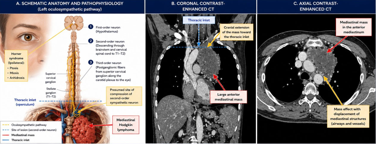
Contrast-enhanced thoracic CT demonstrated a large left anterior mediastinal mass extending into the left pulmonary apex and thoracic inlet, producing mass effect with displacement of adjacent mediastinal structures (Figure 1B and Figure 1C). Its cranial extension toward the thoracic inlet provided a plausible anatomical explanation for compression of the second-order oculosympathetic neuron responsible for the patient’s Horner syndrome.
Brain MRI showed no abnormalities. Angio-CT of the supra-aortic trunks and intracranial vessels revealed a left anterior mediastinal mass with associated laterocervical and supraclavicular lymphadenopathy. Positron emission tomography-computed tomography demonstrated a heterogeneous hypermetabolic mediastinal mass measuring 10 × 6.8 × 7 cm (SUVmax 11.58) with areas of necrosis, along with multiple hypermetabolic supradiaphragmatic lymph nodes.
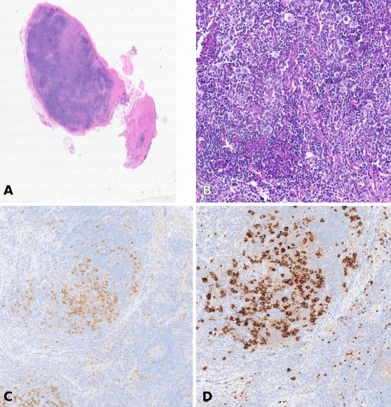
Core needle biopsy followed by excisional biopsy of a retroclavicular lymph node confirmed classical Hodgkin lymphoma, nodular sclerosis subtype. Histopathological examination revealed classic Reed–Sternberg cells within a polymorphous inflammatory background composed predominantly of small lymphocytes, eosinophils, plasma cells, and histiocytes. Immunohistochemistry demonstrated CD30 and CD15 positivity (Figure 2C, Figure 2D), with weak expression of paired box protein 5 (PAX5), CD20 negativity, and negativity for Epstein–Barr virus-encoded small RNAs (EBER).
The patient was diagnosed with classical Hodgkin lymphoma and initiated treatment with the Brentuximab-AVD regimen after sperm cryopreservation.
Discussion
Horner’s syndrome secondary to mediastinal lymphoma is a very rare mode of presentation [4],[5],[6],[7],[8]. The proposed mechanism involves compression or infiltration of the cervical or thoracic sympathetic chain [3]. In our patient, the extension of the mediastinal mass toward the thoracic inlet and pulmonary apex suggests involvement of the second-order oculosympathetic neuron, providing an anatomical explanation for the development of Horner syndrome (Figure 1A, Figure 1B, Figure 1C).
The differential diagnosis of Horner’s syndrome is broad and includes traumatic lesions, carotid artery dissection, brainstem stroke, demyelinating disease, infectious and inflammatory disorders, Pancoast tumors, mediastinal malignancies, and other lesions affecting the sympathetic pathway [1],[2],[3]. Consequently, a systematic diagnostic approach and targeted imaging studies are essential to identify potentially serious underlying etiologies.
In this patient, the history of facial trauma may initially have suggested a traumatic cause; however, persistence of HS prompted further investigation and ultimately led to the diagnosis of mediastinal Hodgkin lymphoma.
Published cases most frequently describe classical Hodgkin lymphoma, nodular sclerosis subtype, occurring in young patients with bulky mediastinal masses [5],[6],[7],[8],[9]. The absence of B symptoms does not preclude the presence of extensive mediastinal Hodgkin lymphoma [9],[10].
This case highlights the importance of excluding mediastinal lesions in any patient with HS not explained by carotid or primary neurological pathology [1],[2], even in the presence of a traumatic history. Positron emission tomography-computed tomography is particularly valuable for staging and therapeutic planning [10].
Conclusion
Horner’s syndrome may represent a sentinel sign of mediastinal Hodgkin lymphoma. Persistence of symptoms warrants comprehensive imaging of the neck and chest to ensure early diagnosis and timely treatment.
LEARNING POINTS
- Horner’s syndrome may be the first manifestation of serious underlying pathology.
- Persistent Horner syndrome warrants imaging of the neck and chest even in patients with a history of trauma.
- Mediastinal masses extending toward the thoracic inlet may compromise the oculosympathetic pathway and cause Horner’s syndrome.
- Hodgkin lymphoma should be included in the differential diagnosis of Horner syndrome, particularly when mediastinal pathology is present or suspected.
REFERENCES
1.
Biousse V, Galetta A, Newman NJ. The clinical spectrum of Horner’s syndrome. Rev Neurol Dis 2004;1(2):45–52.

2.
Kanagalingam S, Miller NR. Horner syndrome: Clinical perspectives. Eye Brain 2015;7:35–46. [CrossRef]
[Pubmed]
3.
Tamhankar MA, Volpe NJ. Neuro-ophthalmic presentation of systemic malignancies. Curr Opin Ophthalmol 2009;20(6):505–12.
4.
Simon SR, Dorighi JA, Branda RF, Ershler WB. Horner’s syndrome: An unusual presentation of Hodgkin’s disease. Med Pediatr Oncol 1985;13(6):390–1. [CrossRef]
[Pubmed]
5.
Micieli A, Ghorab Z, Micieli JA. Horner syndrome secondary to Hodgkin lymphoma. CMAJ 2020;192(8):E187. [CrossRef]
[Pubmed]
6.
Abascal Azanza C, Abarzuza Cortaire R, Plaza Ramos P. Horner’s syndrome: An unusual ophthalmological presentation of Hodgkin lymphoma. [Article in Spanish]. An Sist Sanit Navar 2017;40(3):461–6. [CrossRef]
[Pubmed]
7.
Fernández-Torrón R, Martínez-Vila E. Síndrome de Horner como forma de presentación de linfoma mediastínico: A propósito de un caso. Neurología 2010;25(4):264–7.
8.
Martín-Rodríguez J, de la Iglesia-Martínez A, Brea-Hernando A, Lahuerta-Palacios JJ. Síndrome de Horner como primera manifestación de linfoma mediastinal. Rev Clin Esp 2004;204(3):159–61.
9.
Wakely PE Jr, Geyer JT, Elgert PA. Mediastinal Hodgkin lymphoma: Cytopathologic and immunophenotypic findings. Cancer Cytopathol 2014;122(9):694–702.
10.
Rodriguez FJ, Nasr MR, Inamdar KV, Abdul-Karim FW. Clinical-pathologic features of mediastinal lymphomas. Am J Clin Pathol 2004;121(4):502–12.
SUPPORTING INFORMATION
Author Contributions
Francisco Javier Ruiz Miyares - Acquisition of data, Analysis of data, Revising the work critically for important intellectual content, Final approval of the version to be published, Agree to be accountable for all aspects of the work in ensuring that questions related to the accuracy or integrity of any part of the work are appropriately investigated and resolved.
Franco Emanuel Appiani - Conception of the work, Design of the work, Analysis of data, Revising the work critically for important intellectual content, Final approval of the version to be published, Agree to be accountable for all aspects of the work in ensuring that questions related to the accuracy or integrity of any part of the work are appropriately investigated and resolved.
Fiorella Karina Miraval Márquez - Conception of the work, Design of the work, Acquisition of data, Analysis of data, Drafting the work, Revising the work critically for important intellectual content, Final approval of the version to be published, Agree to be accountable for all aspects of the work in ensuring that questions related to the accuracy or integrity of any part of the work are appropriately investigated and resolved.
Silvia Ruiz De Gracia - Conception of the work, Design of the work, Acquisition of data, Analysis of data, Drafting the work, Revising the work critically for important intellectual content, Final approval of the version to be published, Agree to be accountable for all aspects of the work in ensuring that questions related to the accuracy or integrity of any part of the work are appropriately investigated and resolved.
Antonio Jesús Donaire - Conception of the work, Design of the work, Acquisition of data, Analysis of data, Drafting the work, Revising the work critically for important intellectual content, Final approval of the version to be published, Agree to be accountable for all aspects of the work in ensuring that questions related to the accuracy or integrity of any part of the work are appropriately investigated and resolved.
Guaranter of SubmissionFrancisco Javier Ruiz Miyares, MD, is the guarantor of this submission and accepts full responsibility for the integrity and accuracy of the work.
Source of SupportNone
Consent StatementWritten informed consent was obtained from the patient for publication of this article.
Data AvailabilityAll relevant data are within the paper and its Supporting Information files.
Conflict of InterestAuthors declare no conflict of interest.
Copyright© 2026 Francisco Javier Ruiz Miyares et al. This article is distributed under the terms of Creative Commons Attribution License which permits unrestricted use, distribution and reproduction in any medium provided the original author(s) and original publisher are properly credited. Please see the copyright policy on the journal website for more information.